COVID-19
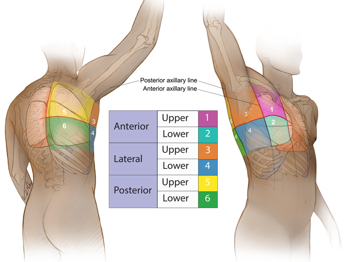
- Indicator up towards the head
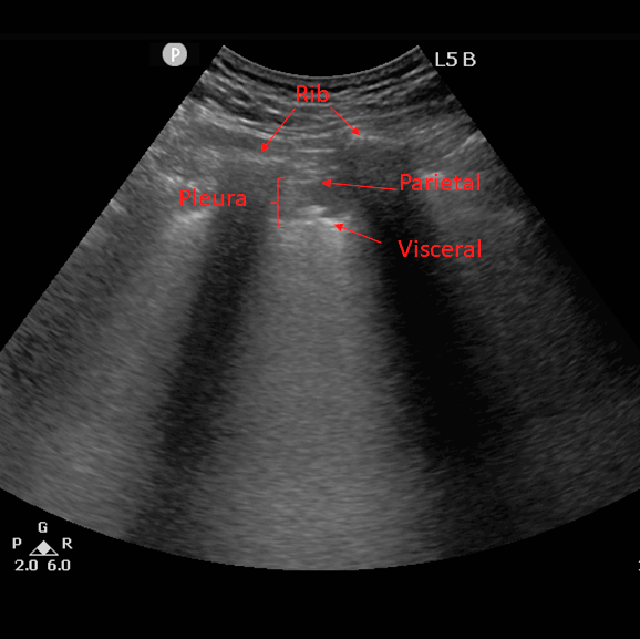
- Place over a rib, identify pleural line
- Image each of the 6 zones with a LF AND HF transducer
- Left and then Right side
- Label side, Zone, Transducer (i.e. R2 LF)
- Image Zones the same way each time (Left Zone 1,2,3,4,5,6 and Right Zone 1,2,3,4,5,6)
- If patient being proned a)Anterior and Side (L 1,2,3,4 then R 1,2,3,4) and then b)Posterior (L 5,6 then R 5,6) when patient flipped
- Completely image ALL of each zone
- If pathology save 3-5 second clip of area if interest
- If no pathology still save clip
- Best seen with a low frequency probe
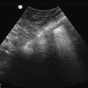
- Focal B-lines early (Progressive to confluence, Multi-lobar distribution, and Bilateral distribution)
- Pleural effusions are rare
- A-lines during recovery
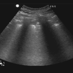
- Best visualized with a high frequency probe
- Identify the pleural line and then rotate the probe horizontally (thickened, irregular pleural line, sub pleural consolidations, and localized pleural effusion adjacent to consolidations)
- Always save a clip of each zone
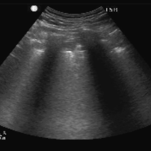
- Curvilinear allows seeing more rib spaces
- B lines in a variety of patterns including focal, multifocal, and confluent
- Consolidations in a variety of patterns including multifocal small, non-translobar, and translobar with occasional mobile air bronchograms
- Appearance of A lines during recovery phase
- Pleural effusions are uncommon
Figure 2 - B-lines in COVID-19 Pneumonia
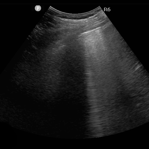
Figure 3 - Pleural Findings in COVID-19 Pneumonia (High Frequency)
Figure 4 - Pleural Findings in COVID-19 Pneumonia (Curvilinear)
Clip 1 - Conflucence of B-lines
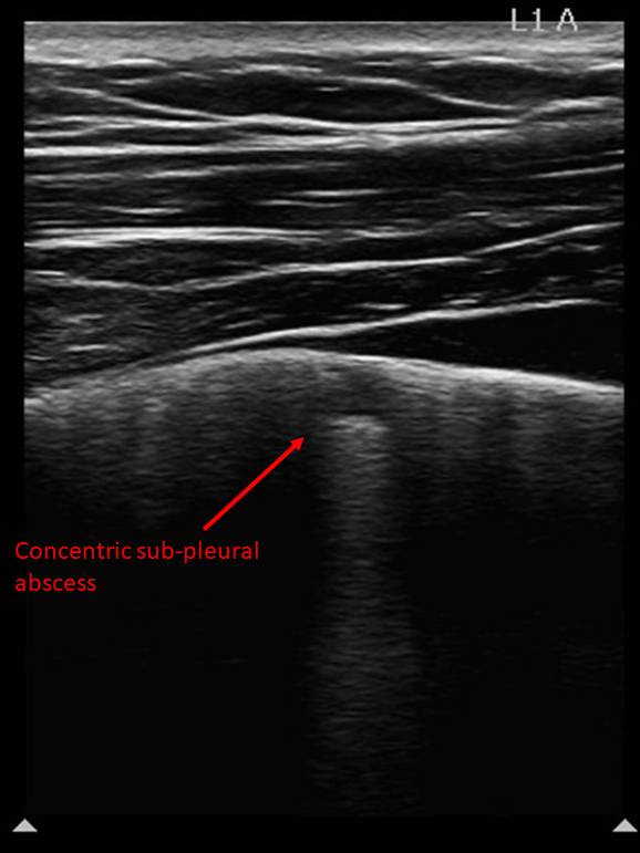
Clip 2 - Subpleural Abscess (High Frequency)
Clip 3 - Subpleural Abscess (Curvilinear)