Ejection Fraction
- Visual assessment of all 4 windows, will get a good sense from the PLA.
- Look for thickening of LV walls during systole and not just the swinging motion of heart.
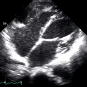
- Also look for longitudinal movement of mitral annulus in the A4C view.
- Estimate the percent (%) ejected with each heart beat.
- Slow the video down and look at the LV at end diastole and end systole; at its largest and at its smallest cavity size.
- Mentally trace the inside of the LV at end diastole and end systole.
- Not as hard as it seems, and more reproducible the most measurements.
EF > 70% → Hyperdynamic: Excellent function, complete ejection, LV empty at end systole
EF 55-70% → Normal: Good function, most blood is ejected, some blood remains at end systole
EF <55, >40% → Mild LV dysfunction: Decreased function, <55% of blood is ejected, blood remains at end systole
EF <40, >30% → Moderate dysfunction: Very dysfunctional, little blood is ejected, blood remains at end systole
EF <30% → Severe dysfunction: Barely moving, almost no blood is ejected, full at end systole
Do not overthink; it will become intuitive quickly.
Think of it is as: Excellent, Good, not as good as I want, poor, and shockingly bad.
Foreshortening of ventricles will result in overestimation of EF.
Heart rate effect may make EF falsely look better (w/ tachycardia) or worse (w/ bradycardia) in some patients.
Most errors occur between mildly dysfunctional and lower end of normal.
Do not worry too much about this difference, ICU patients should have a high EF. Low end of normal and mildly dysfunctional are both problematic.
Make sure to use cardiac exam or presets. If using abdominal imaging the heart will look dysfunctional.
Clip 1 - Normal LV EF, parasternal long view
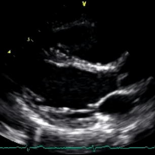
Clip 2 - Severe LV dysfunction, parasternal long view
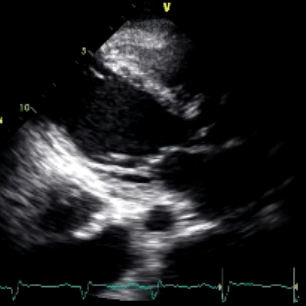
Clip 3 - Normal LV EF, parasternal short view
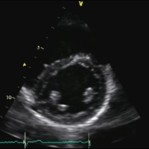
Clip 4 - Severe LV dysfunction, parasternal short view
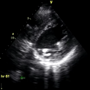
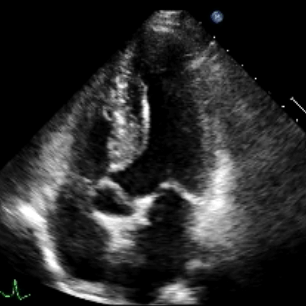
Clip 5 - Normal LV EF, apical 4 chamber view