2D and B-mode
Two dimensional (2D) and B-mode refer to a moving clip created by scan lines sequentially sent through a field at a frame rate. It is the most common mode of imaging, and is excellent for global assessments of structure and function.
- Select 2D or B-mode
- Adjust the time for longer clips
- 5 heart beats or 3-5 seconds
- Allows better assessment of function
- Save clips
- Needed for quality improvement, teaching, and billing
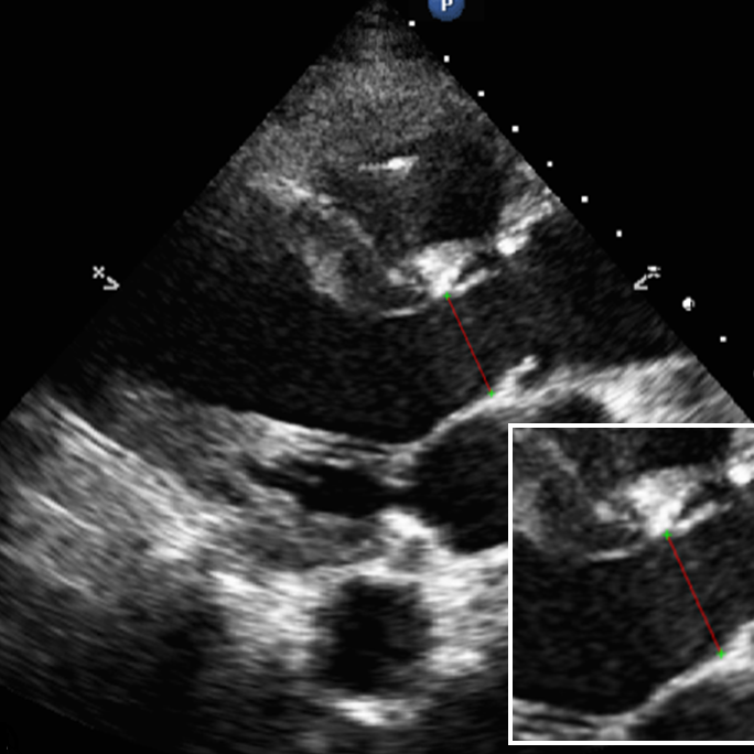
- Calpier is a tool for measuring structures
- Left ventricular outflow tract diameter
- IVC diameter and diameter change
- Optic nerve sheath diameter
- Image area of interest in 2D
- Use active zoom if available
- Save video clip
- Freeze image
- Measure edge to edge (Figure 1)
- Save measurement
Most systems will have an auto-optimize button (different names depending on the manufacturer)