Lung B Lines
- Use a phased array ultrasound probe in abdominal mode.
- Place the indicator up towards the head and place the probe on a selected intercostal space.
- Obtain views of the upper, middle and lower sections of the lungs bilaterally.
- Try to do it the same way every time (LU, LM, LL and then RU, RM. RL) to make interpretation easier.
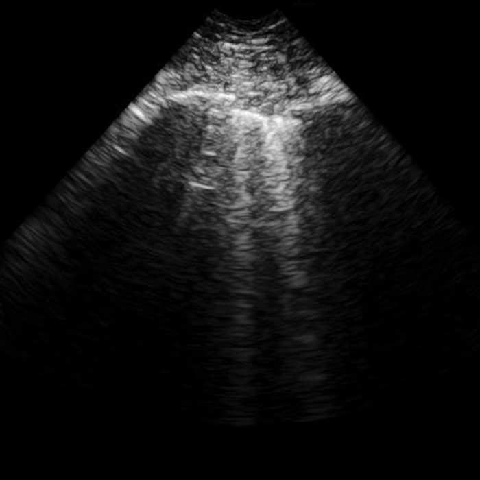
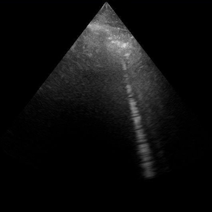
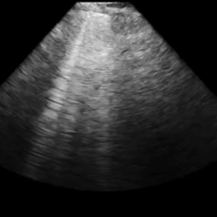
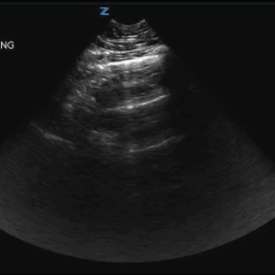
- B-lines are discrete hyperechoic reverberation artifacts that originate from the pleural line and extends towards the bottom of the ultrasound screen.
Evidence of 3 or more B-Lines in multiple intercostal spaces suggests lung interstitial fluid.
Healthy individuals can have B-Lines present in the lower lung portions.
Lung interstitial fluid can be caused by pulmonary edema of various causes, interstitial pneumonia or pneumonitis and diffuse parenchymal lung disease (ie pulmonary fibrosis).