RUSH
RUSH stands for “Rapid Ultrasound for Shock and Hypotension”. It is designed to be a quick and easy to perform. The primary indication is undifferentiated hypotension.
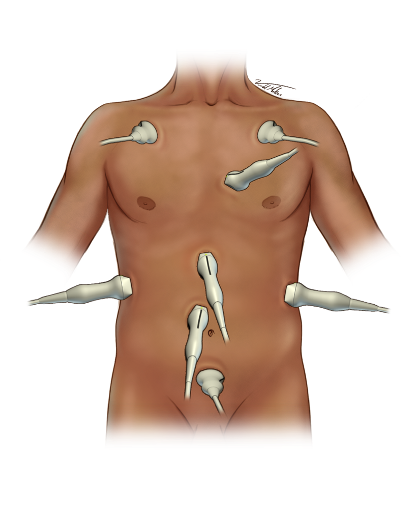
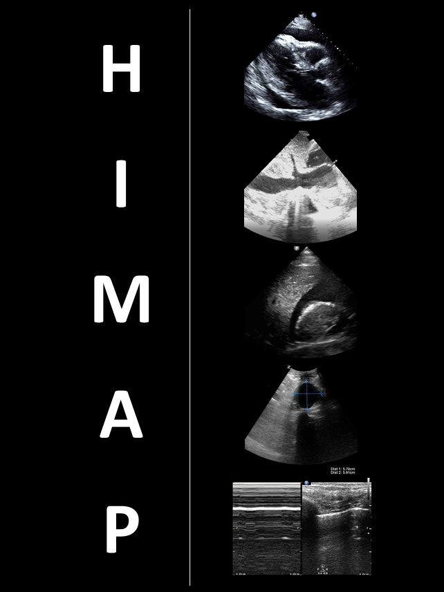
Use a curvilinear probe with the abdominal preset exam mode to obtain the windows (Figure 1). The components of the exam can be memorized using the HI-MAP mnemonic (Figure 2).
HI-MAP = Heart IVC Morrison’s pouch Aorta Pneumothorax
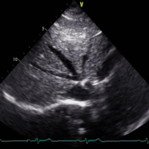
- Assess for pericardial fluid and tamponade (Clip 1)
- Assess RV function (Note apical sparing and other findings consistent with pulmonary embolus)
- Assess LV function
- Long axis view
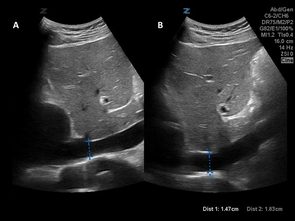
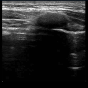
- Assess IVC diameter
- Assess IVC diameter change
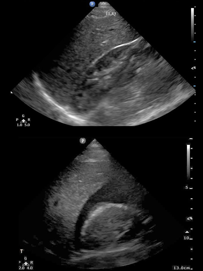
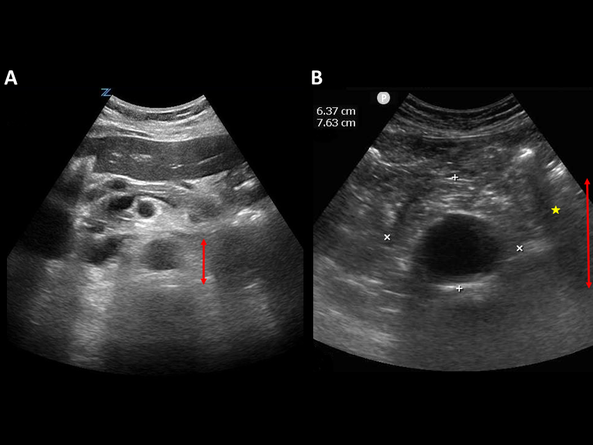
- Assess free fluid within Morrison’s pouch
- Extend to full FAST when indicated
- Transducer below SX
- Indicator to patient’s right
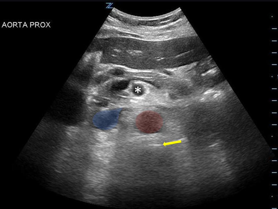
- Aorta is circular structure right of the IVC (Figure 5)
- Scan down to from the celiac branch to the bifurcation
- Measure upper outer wall to just above vertebral body (Figure 6)
- Larger than 5 cm is abnormal, increase risk of rupture
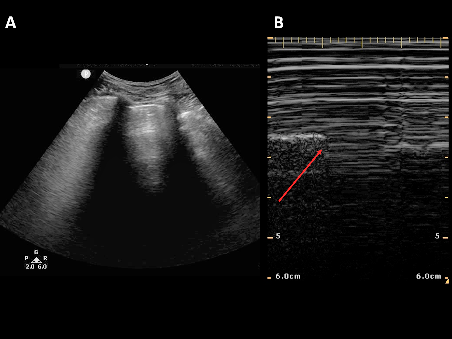
- Bi-lateral 2nd and 4th intercostal space
- Assess lung sliding
- Look for lung point
Figure 4 - Morrison’s Pouch/FAST Windows
Figure 6 - Measuring Aorta and IVC