Subxiphoid
The ideal subxiphoid view includes all four chambers of the heart. The SX should be performed in cardiac presets, but for the RUSH and FAST it is done in abdominal presets to allow rapid imaging of both abdominal and heart in an unstable patient.
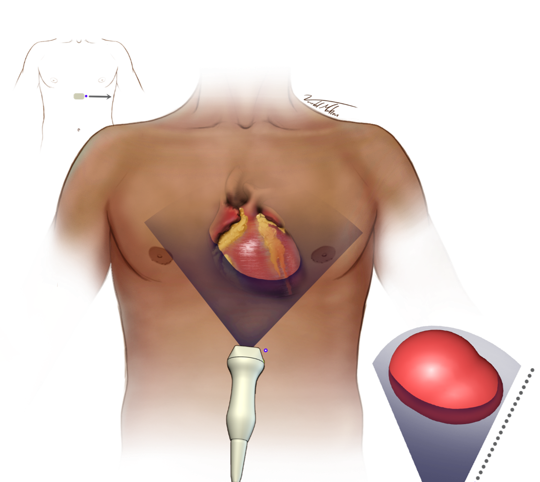
- Overhand grip
- Transducer 2 cm below the xiphoid process
- Directed underneath the ribcage, towards the patient’s left shoulder (2 o-clock position, angle typically less the 20
- May need to increase depth to 20 cm
- Slightly rock the transducer under the sternum, and once under the sternum tilt the face up to open the left ventricle
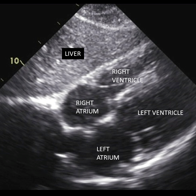
- The left hepatic lobe will be viewed in the near field of the image (Figure 3)
- The subxiphoid four chamber view of the heart can be found deep to the liver (indicator on the right of the screen)
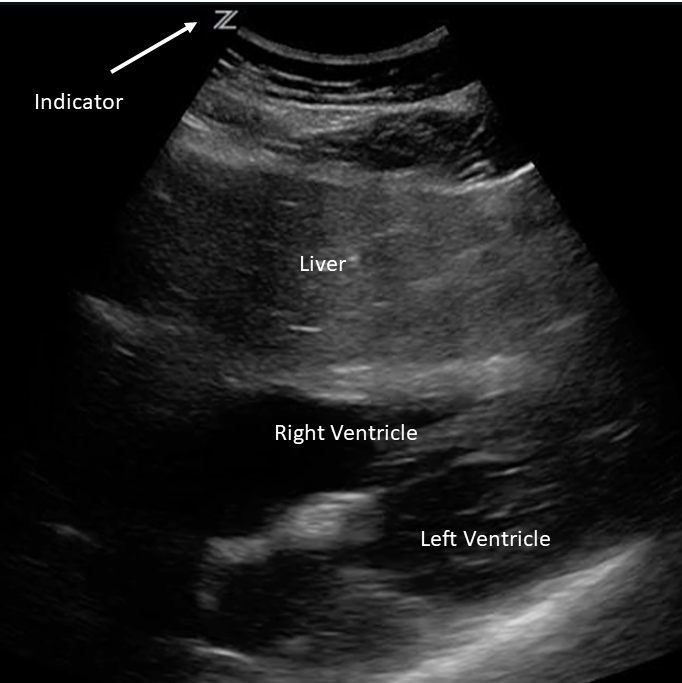
- Abdominal presets are ideal for visceral imaging, not the rapidly moving heart. The liver will appear smoother and more resolved then it does in cardiac presets. But the moving heart will appear less clear and may look dysfunctional. The FAST and RUSH use abdominal presets to allow rapid imaging of both the heart and abdomen. If a more complete cardiac assessment is needed, then cardiac presets should be used.
- Transducer grove is towards the patient’s right (indicator on the left side of the screen)
This view allows global assessment of LV and RV functions
Figure 1 - Transducer Placement
Figure 3 - Ideal Subxiphoid - Cardiac Preset
Figure 4 - Ideal Subxiphoid - Abdominal Preset
Clip 1 - Subxiphoid with Cardiac Preset