M-mode
- M-Mode is Time Motion Display. This images the movement of structures over time.
- Ice pick view through a 2D image
- Single crystal sends and receives a signal, so no frame rate
- Ideal for precise measurements of change over time
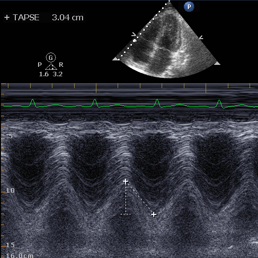
- Tricuspid Annular Plane Systolic Excursion (TAPSE)
- E-Point Septal Separation (EPSS)
- Respiratory change in the Inferior Vena Cava (Delta IVC)
- Locate area of interest in 2D
- Select M-mode to activate cursor
- Place cursor on region to be measured (Video 1)
- Select M-mode again to obtain image (Figure 1)
- Freeze loop, scroll to find ideal point for measurement
- Make measurement, and save image
Figure 1 - Measuering TAPSE with M-Mode