Renal Vein Flow
- Use a phased array transducer in abdominal mode.
- Position the probe in the anterior axillary line by the 11th or 12th rib.
- Keep the transducer groove up and scan posteriorly until kidney comes into view.
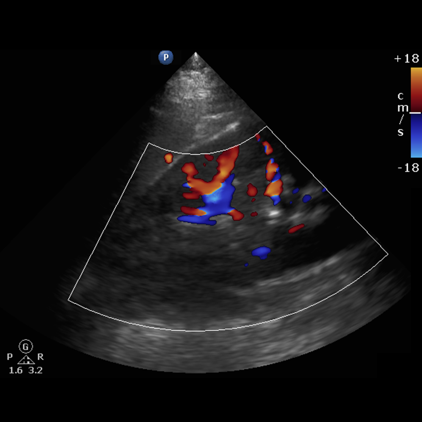
- Place the color Doppler field over the cortex of the kidney.
- Set the Doppler scale to visualize the renal vasculature.
- Note that the abdominal scale usually starts 18 cm/sec, may need to adjust.
- If unable to see anyflow at 10 cm/sec likely not obtainable.
- Put pulse Wave (PW) Doppler over the renal vasculature.
- Reduce gate as low as possible.
- Obtain waveform.
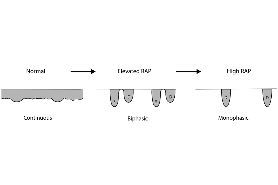
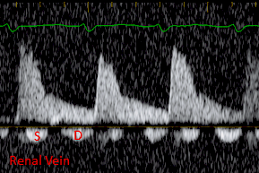
The normal renal vein flow is a continuous waveform.
As RAP increases the waveform changes from continuous to pulsatile.
Pulsatile flow still has some flow throughout the cardiac cycle.
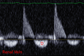
RAP becomes elevated pulsatile flow becomes biphasic (S>D) and then (D>S), progressing monophasic (D only).
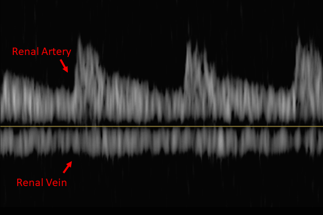
Obtaining the renal artery at the same time helps identify systole and diastole.
Can be difficult to visualize, attempt right side first, move to left if flow cannot be seen.
Prop patient right side up slightly if needed.
Adjust gain until a good color flow signal can be seen.
Often the arterial and venous flow can be seen at the same time.
Figure 1 - Renal Vein Color Doppler
Figure 2 - Normal Renal Vein PW Doppler
Figure 3 - Renal Vein Flow Changes with Rising RAP
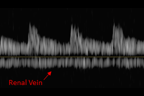
Figure 4 - Renal Vein, Normal PW Doppler
Figure 5 - Renal Vein, Biphasic PW Doppler Pattern
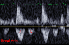
Figure 6 - Renal Vein, Biphasic PW Doppler Pattern
Figure 7 - Renal Vein, Monophasic PW Doppler Pattern