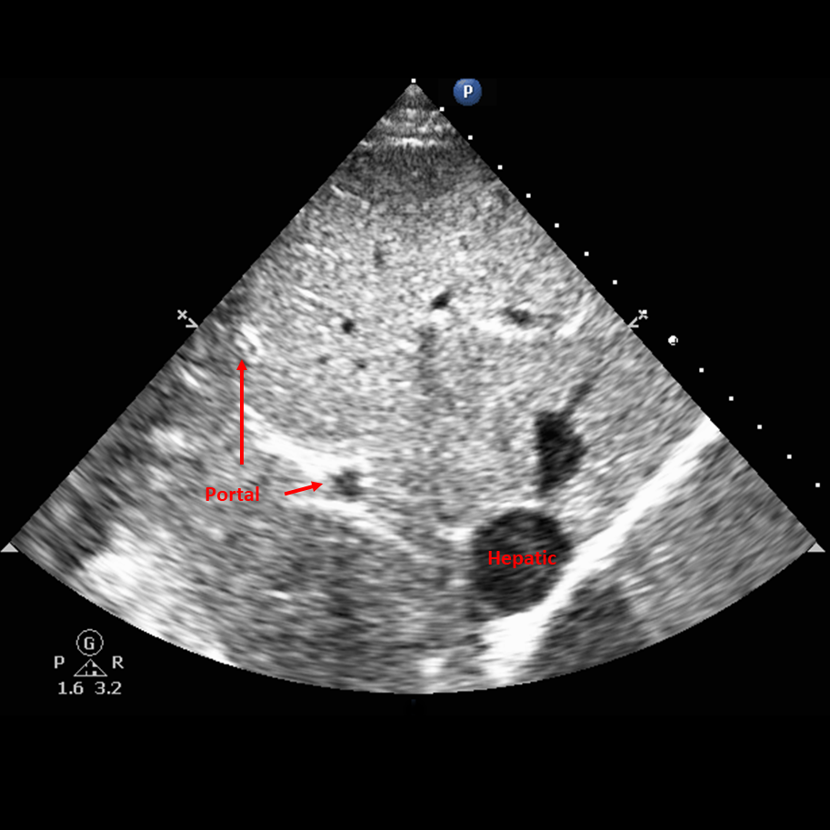
Portal Vein Flow
- Use the phased array probe in cardiac presets.
- Anterior axillary line with the pointer towards the patient’s head.
- Identify the portal vein by the hyperechoic walls (Figure 1).
- Place the color Doppler over the portal vein to identify flow.
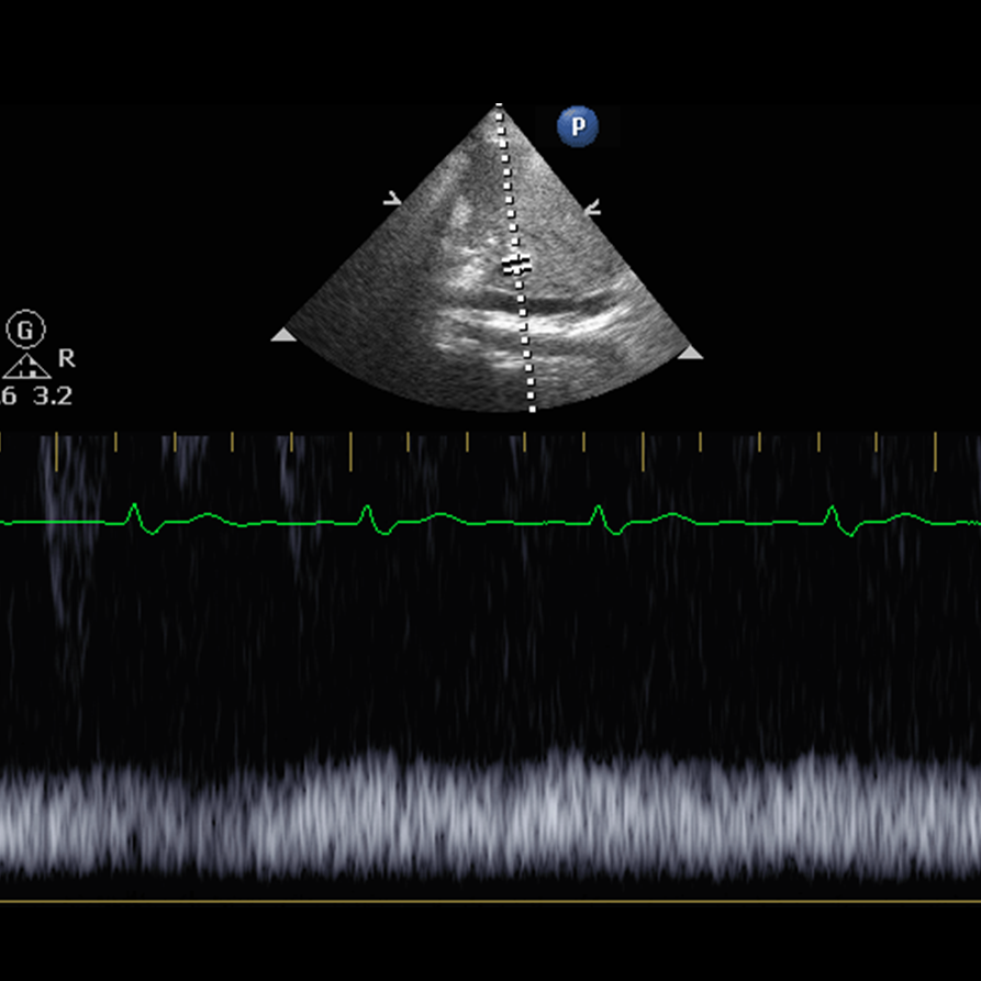
- Place the pulse wave Doppler over the portal vein.
- Reduce the gate as low as possible.
- Acquire PW signal.
- Adjust the scale to optimize waveform.
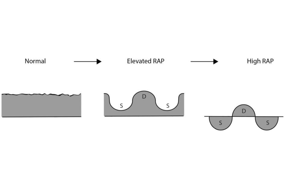
Waveform (Figure 2)
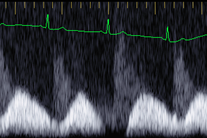
Normal portal flow is continuous. (Figure 3)
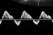
Elevated right atrial pressure (RAP) will cause pulsatility in the vein (Figure 4).
As it increases elevated RAP will cause a to-and-fro pattern (Figure 5).
It is important to differentiate hepatic veins from portal veins. Hepatic veins have hypoechoic thin walls.
Pulsatile flow is also a sign of portal hypertension, and cannot be used as a measure of right atrial pressure elevation in cirrhosis.
Figure 2 - Portal flow patterns with RAP