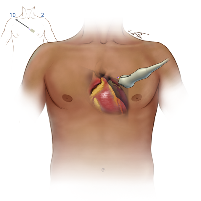
Parasternal Long
- Place the transducer to the left of the sternum
- The indicator bisects the patient’s right clavicle (10 o’clock position)
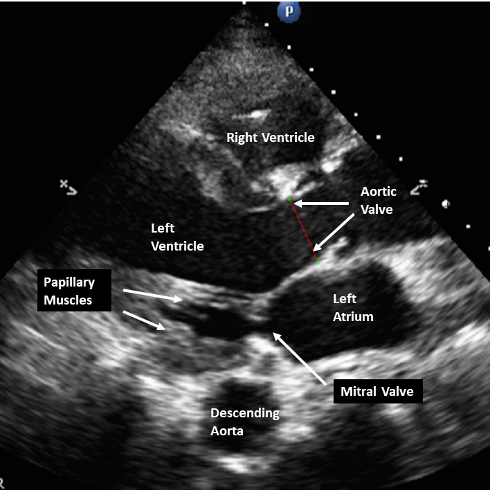
- Slightly rock the transducer under the sternum and once under the sternum tilt the face up to open the left ventricle
- Cardiac exams the indicator is displayed on the right of the screen. Note that in abdominal and all other ultrasound imaging it is displayed on the left
- The indicator is on the imager’s left but displayed on the right of the screen
- The left atrium, left ventricle, left atrium, aortic and mitral valves are will visualized
- LV ejection fraction (visual assessment and E-point septal separation)
- Aortic valve (aortic stenosis and aortic insufficiency)
- Left ventricular outflow tract diameter (needed for stroke volume and cardiac output measurements)
- Left atrial size
- Mitral valve function
- RV function
Start at the 2-6th rib space, but may be lower and more medial in patients on a ventilator.
Figure 1 - PLA Transducer Placement