E-Point Septal Separation
Obtain a PLAX view with visualization of the mitral valve leaflets and interventricular septum.
Select M-mode and place the cursor at the tip of the anterior mitral valve leaflet.
Measure the distance from the interventricular septum to the point of maximal excursion of the anterior mitral valve leaflet (E-wave) on the m-mode tracing.
Normal: <7mm
Mild/Moderate LV Dysfunction (EF 30- 50%): 7-13mm
Severe LV dysfunction (EF <30%): >13mm
The more horizontal the ventricles lie in PLAX the more accurate the measurement.
Place the M-mode cursor as close to the tip of the mitral valve leaflet as possible.
Measurement not accurate in patients who have pathology of the mitral valve or moderate to severe aortic regurgitation.
Patients with severely thickened interventricular septum such as in LVH or HOCM can have an overestimation of LV function.
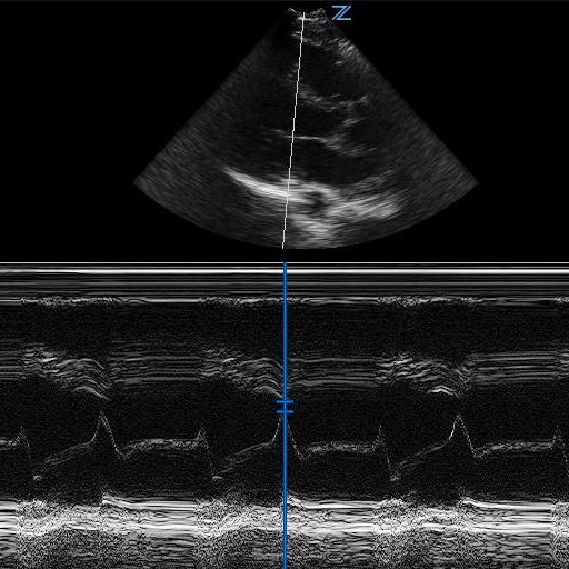
Figure 1 - EPSS with normal LV function
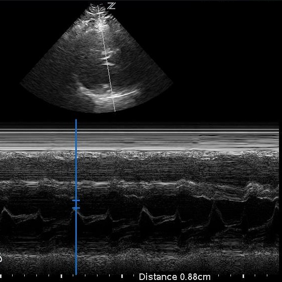
Figure 2 - EPSS with low LV EF