Stroke Volume
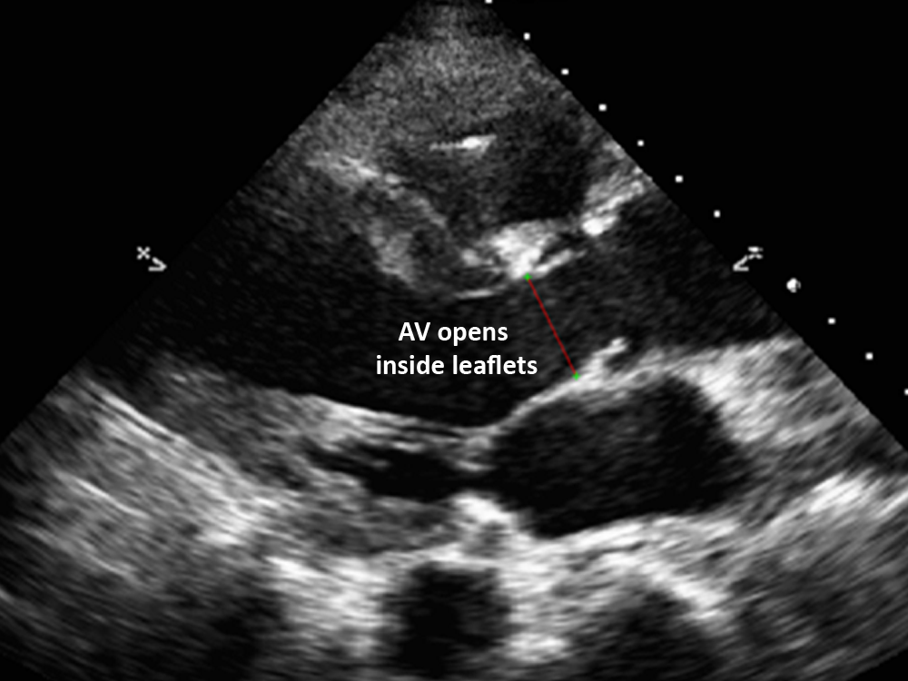
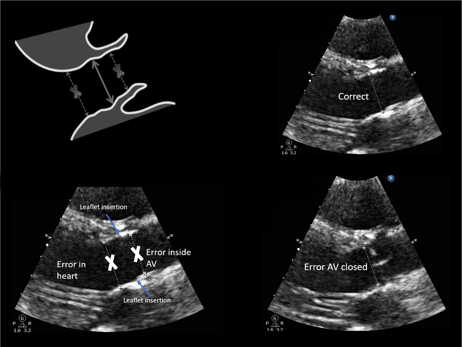
Stroke volume is calculated from two measurements: Left ventricular outflow tract diameter (LVOTd):
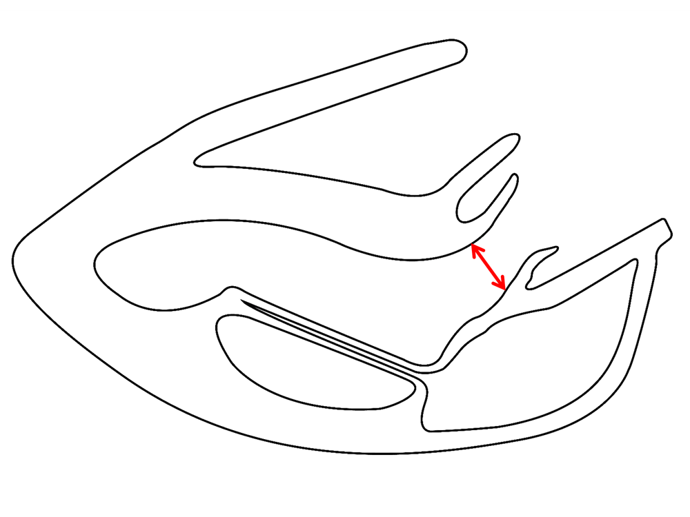
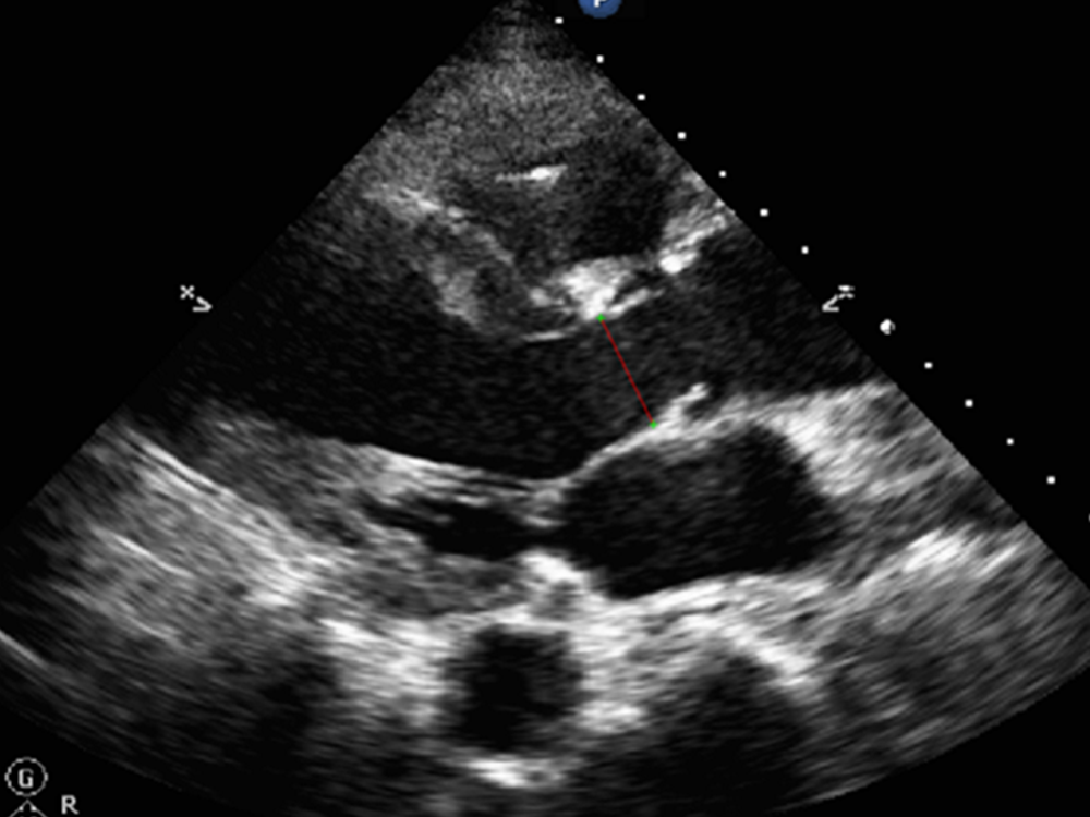
- Measured from the PLA in mid- systole, with active zoom if available.
- Measured from inner edge to inner edge before the bump of leaflet insertion on LV side.
- Normal 1.8-2.2 cm.
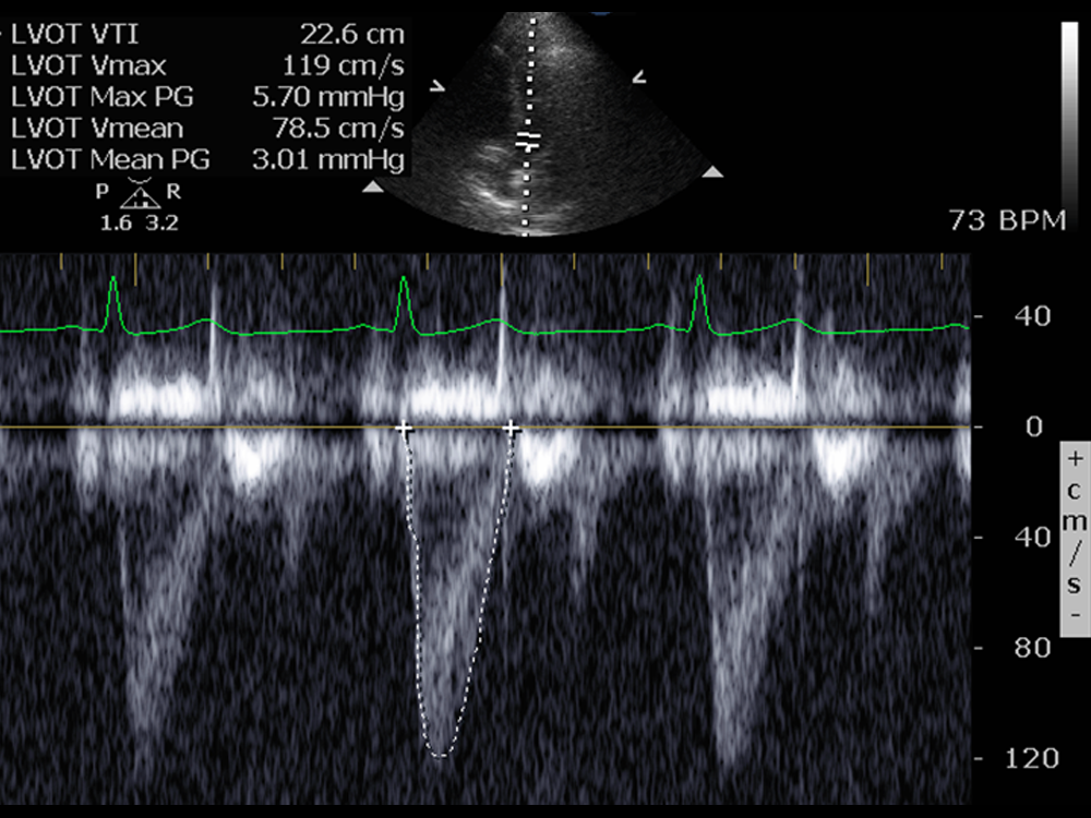
Left ventricular outflow tract velocity time integral (LVOT VTI):
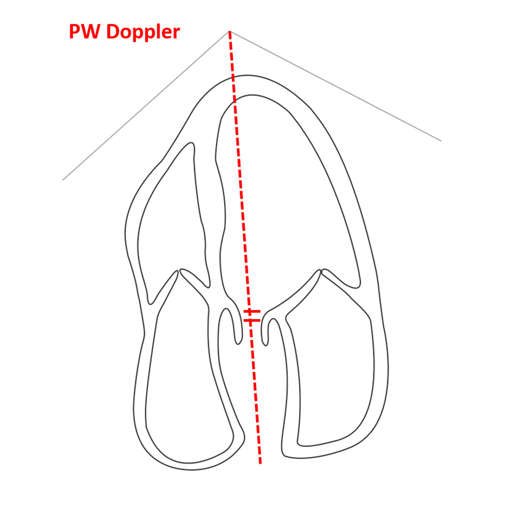
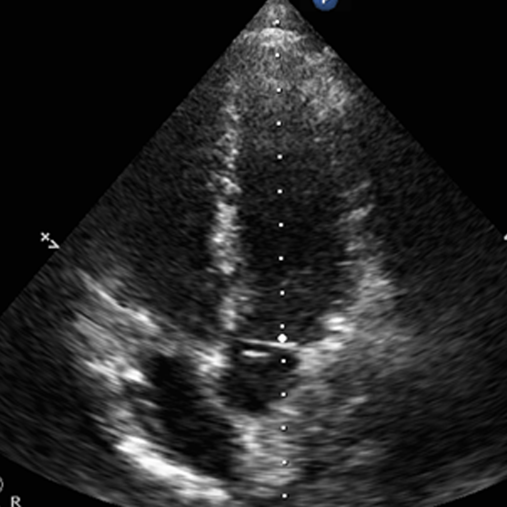
- Measured from the AP 5.
- Place the pulsed wave doppler cursor just before the AV leaflets.
- Trace the envelope of flow, make sure to start and end at the baseline, from when the valve opens til it closes.
- Normal 16-35 cm.
Calculated using the following formula: SV = π x (LVOTd/2)2 x LVOTVTI
- Look closely at the aortic valve for stenosis.
- Related to BSA smaller people <1.8 cm and larger >2.2 cm.
- Anatomic fact does not change rapidly in an adult, so only needs to be measured once in an adult.
- Measured from the AP 5.
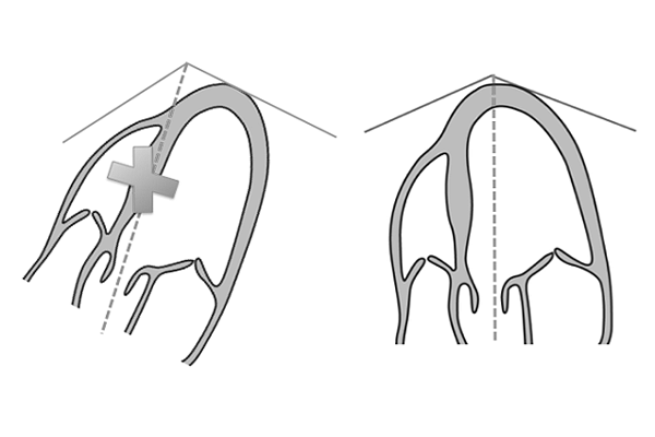
- Do not count if angle > 15% from midline.
- Do not count if passes through septum.
- Select largest envelope if heart beat regular or average of 3-5 beats if irregular.
- Velocity can be underestimated with aliasing.
- LVOT VTI is not a reliable tool in the presence of aortic valve stenosis.
Figure 1a - how to measure LVOTd
Figure 1b - how to measure LVOTd
Figure 2a - how to measure LV VTI
Figure 2b - how to measure LV VTI
Figure 3 - correct measurement of LVOT VTI
Figure 4 - correct measurement of LVOTd
Figure 5 - erros in measurement of LVOTd
Figure 6 - correct cursor placement for measuring LVOT VTI
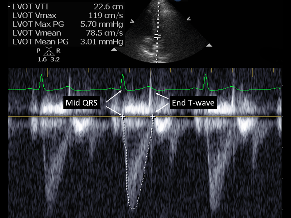
Figure 7 - using the ECG to measure the LV VTI
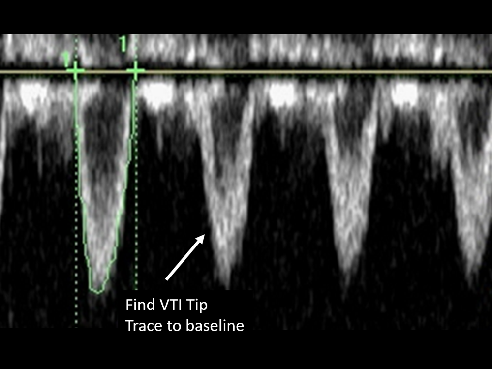
Figure 8 - tracing the LV VTI without the ECG
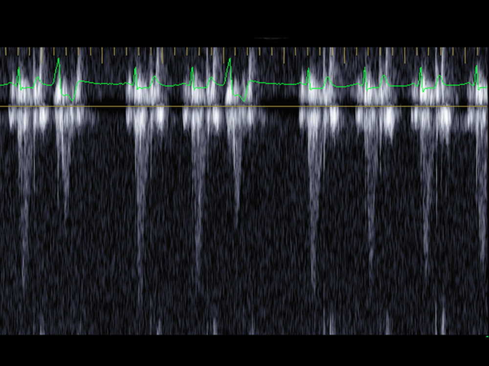
Figure 9a - poor LV VTI signal
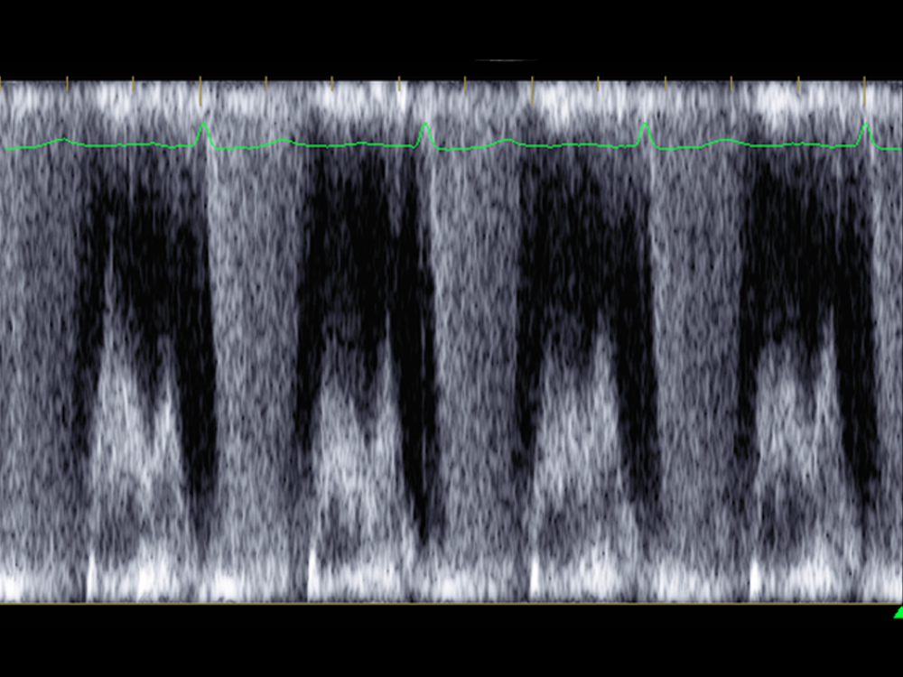
Figure 9b - poor LV VTI signal
American Society of Echocardiography Guidlines: Assessment of Structure and Function.