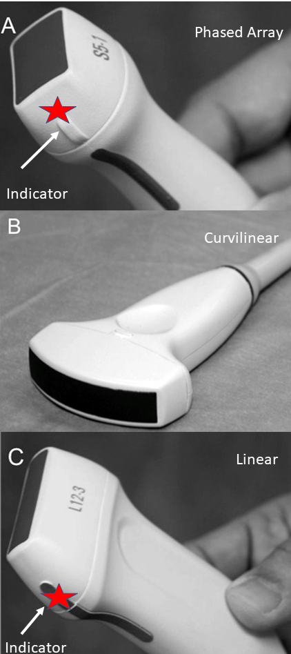
Probe Selection
- Cardiac imaging
- Focused Assessment Sonogram for Trauma (FAST)
- Deep Lung imaging
- Abdominal and pelvic imaging
- Aortic imaging
- Deep Lung imaging
- FAST
- Rapid Ultrasound in Shock (RUSH)
- Vascular access
- Detailed Lung imaging
- Superficial imaging
- Presets and Exam Type are preset features which optimize different types of imaging
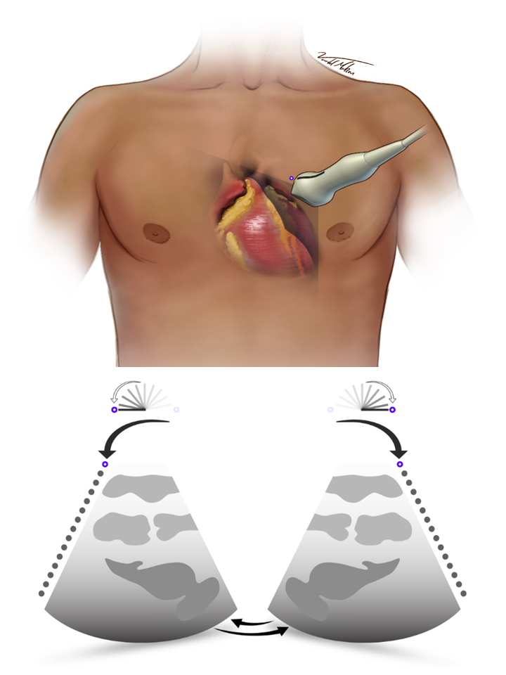
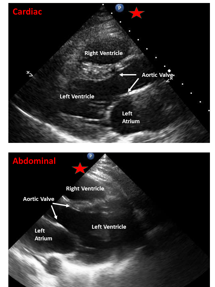
- We strongly recommend performing heart sonograms in cardiac presets
- Except for the subxiphoid window in the FAST and RUSH, combine abdominal and cardiac imaging in a rapid assessment
- Optimized for assessing the rapidly moving heart
- Transducer indicator is displayed on the right of the screen (by convention/historical, and possibly to correlate with cardiac catheterization images)
- Faster frame rate with less scan lines (grainy still image, but better moving image)
- Less compression (appears more black and white)
- Optimized deep imaging of viscera, less movement the heart
- Transducer is displayed on the left of the screen (most ultrasound imaging displays on the left)
- Lower frame rate with more scan lines (better images of still structures such as the Liver, but heart can appear dysfunctional)
- More compression (Smooth grey images)
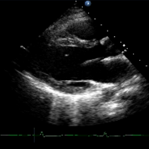
Figure 3 - Abdominal and Cardiac Views
Clip 1 - PLA with Abdominal Preset