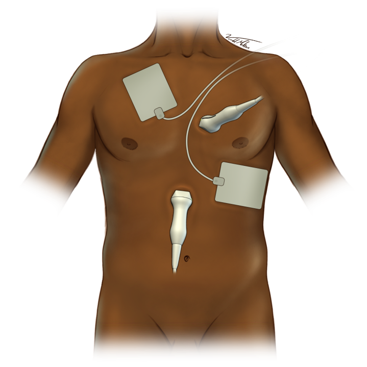
CASA
The Primary Indication is in Cardiac Arrest. Performance of US must not interfere with CPR.
- Assess cardiac activity
- Assess pericardial effusion/tamponade
- Assess right heart strain (possible PE)
- Rule out large pneumothorax
- Assess for intra-abdominal hemorrhage (FAST)
- Read/evaluate saved clip
- Assess cardiac activity
- Record 10 second clip
In non-shockable rhythms, POCUS can identify reversible causes of CA, such as tamponade, pulmonaryembolism, hypovolemia and tension pneumothorax.
- Differentiate a true asystole (cardiac standstill) versus a still-contracting heart.
- Patients with cardiac contracility have a higher chance of achieving ROSC.
Phased Array in cardiac presets/exam type
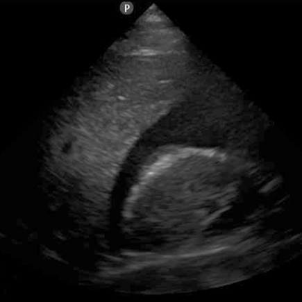
- Rapidly assess for cardiac activity (Clip 1)
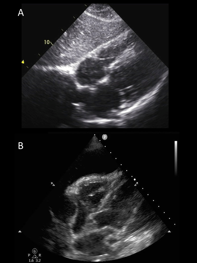
- Assess for pericardial fluid/tamponade (Figure 2)
- Assess RV function/size
- If unable to obtain, parasternal long axis (PLA)
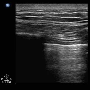
- Assess lung sliding right and left of the sternum
- Assess for free fluid (possible hemorrhage)
POCUS must not interfere with the CPR
ROSC should be determined based on the presence of a pulse, capnography and arterial pressure readings
Figure 2 - (A) Normal Pericardium (B) Cardiac Tamponade